
Skip to content

One RIS/PACS for the entire life of a study

RIS/PACS platform
From patient admission in the HIS to the signed, published report, over open standards — HL7, FHIR, MWL and DICOM — in a single traceable workflow.
End-to-end workflow
Clinical flow and acquisitionResult distribution
NOVA Imaging · RIS/PACSHL7 · FHIR · ORUMWLDICOMBillingResultsRISOrchestratorHIS / EMRPatient · visitModalitiesCT · MR · US · DXPACSDICOM archiveReadingViewer · reportERPBillingPortalsPatients · physicians
WorklistReading
Prioritize critical
ModalitiesPrioritiesPayersCreate list
All7My studies0Mammography0Columns
ModPatient · studyPrioTime
DX
Urgent
Gómez M., J.C.
Chest PA · EX-0001
14m
MR
Rodríguez P., M.
Brain MRI · MR-0156
HIGH2d
US
López S., C.
Renal ultrasound · US-0234
HIGH2d
CT
Díaz G., F.
Abdominal CT · CT-0456
MED5d
MG
Torres N., P.
Bilateral mammogram · MG-0678
LOW7d
7 pending · view "All"Shared · night shift

Intelligent worklist

Prioritizes studies by clinical urgency and SLA, with filters by modality, payer contract and status.

  • Automatic prioritization by urgency
  • Filters by modality, payer contract and site
  • Alerts for breached or near-breach SLAs
portal.novaimaging.co/viewer
Synced
RID · XXXXXXX
CT CHEST-ABDOMEN
01-Jul-2026 · 02:19:12
AW:360 L:60181/362
AID · XXXXXXX
CT CHEST-ABDOMEN
01-Jul-2026 · 02:19:12
HW:360 L:60253/512
RID · XXXXXXX
CT CHEST-ABDOMEN
01-Jul-2026 · 02:19:12
HW:360 L:60257/512
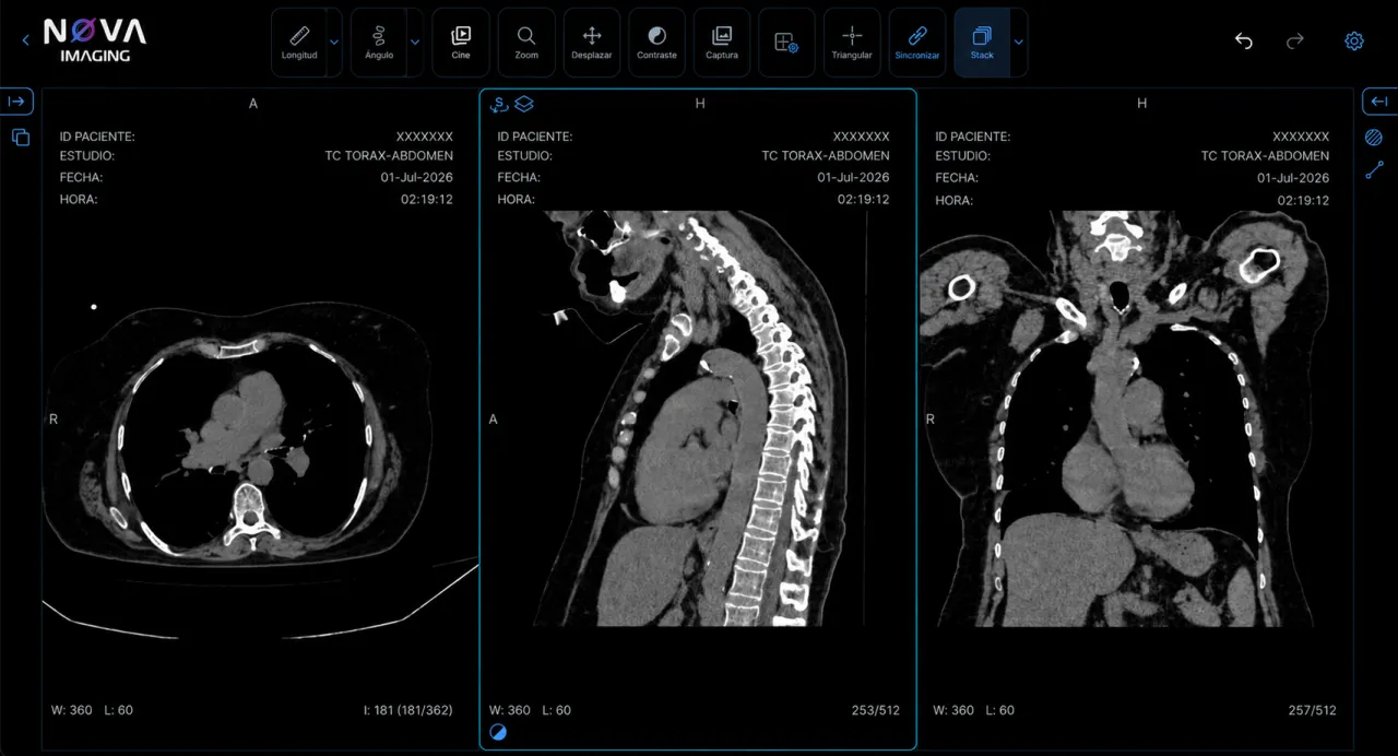
NOVA Imaging viewer: chest-abdomen CT in axial, sagittal and coronal planes

Native DICOM viewer

Fluid reading with measurement tools, window/level and comparison series — no plugins.

  • Measurement, window/level and annotations
  • Comparison of series and prior studies
  • No plugins to install on the workstation
report editor · AI
Findings

Focal lesion in the left kidney,

of cystic appearance, measuring 42 mm

with well-defined borders.

AI detectionLive
Laterality

“Left” does not match the R marker on the image.

Classification

Renal cystic lesion with no Bosniak category assigned.

Structured reporting

Templates per study type, with macros and digital signature.

  • Templates configurable per modality
  • Macros and predefined phrases
  • Digital signature with traceability
HIS / EMRMedical recordPatient portalStudies and reportsPhysician portalReferrersERPBillingNOVA Enterprise ImagingFHIR · HL7 · DICOM

HL7/FHIR interoperability

NOVA Imaging connects with your HIS, ERP and portals over open healthcare standards — without replacing the systems you already use.

  • HL7 v2 (ADT · ORM · ORU) and FHIR R4 over a REST API
  • DICOM and DICOMweb (WADO · QIDO · STOW), MWL and IHE profiles (XDS-I · PIX/PDQ · SWF)
  • Standard clinical terminologies: LOINC, SNOMED CT and ICD-10
  • Orders, results and statuses synced with the HIS and ERP in real time
portal.novaimaging.co
EMElena MarínPatient portal · 3 studies
MRRight knee MRIJul 12, 2026View study
CTAbdominal CTMar 3, 2026View study
USThyroid ultrasoundNov 18, 2025View study

Patient portal

Results and images delivered online, with secure access.

  • Access through a secure, controlled link
  • The patient’s study history
  • Report and image download
portal.novaimaging.co/viewer
Synced
RID · XXXXXXX
CT CHEST-ABDOMEN
01-Jul-2026 · 02:19:12
AW:360 L:60181/362
AID · XXXXXXX
CT CHEST-ABDOMEN
01-Jul-2026 · 02:19:12
HW:360 L:60253/512
RID · XXXXXXX
CT CHEST-ABDOMEN
01-Jul-2026 · 02:19:12
HW:360 L:60257/512
NOVA Imaging viewer: chest-abdomen CT in axial, sagittal and coronal planes

Zero-footprint visualization

Complete DICOM series from any browser, with no client to install.

  • Ideal for remote tumor boards
  • Second opinions with nothing to install
  • Works on any machine with a browser
quality dashboard
Risk level
Low10%Medium72.5%High17.5%
By modality
CTUSMREPDX
Summary by physician
M. Torres Vega98.9Excellent
R. Salas Nieto100Excellent

AI report quality control

Automatic review of signed reports against the findings in the study.

  • Compares the report text against the viewer
  • Flags omissions before delivery
  • Reduces the risk of human error

Real-time inconsistency detection

Alerts while the radiologist is writing the report.

  • Checks as the report is written
  • Alerts on laterality mismatches
  • Fewer reworks after signing
Visualization

Diagnostic-grade visualization, in the browser

The NOVA Imaging viewer is built on the reference open-source ecosystem for web imaging (Cornerstone3D · OHIF), with a GPU rendering engine and multi-modality support — with nothing to install on the workstation.

Fully web-based viewer (zero-footprint)

It opens in the browser and streams pixels on demand, so large studies load in seconds with no client to install.

GPU-accelerated 3D rendering

A Cornerstone3D engine with GPU acceleration: scroll through heavy series and 3D volumes fluidly.

Multiplanar reconstruction (MPR)

Synchronized axial, coronal and sagittal slices, with annotation navigation across planes.

2D and 3D segmentation

Labelmaps and surfaces in 2D and 3D to mark structures and measure on the volume.

Measurements, annotations and SR

Length, angle, ROI, bidirectional and more, with measurement tracking and structured reporting (DICOM SR).

Hanging protocols and comparison

Configurable hanging protocols and comparison with prior studies, to read faster and with context.

Also supports:PET/CT fusion4D / dynamicVideoMicroscopy (WSI)RT Structures and contouringUltrasound modeMammographyPDF
Infrastructure and storage

Intelligent cloud storage, with no hidden costs

NOVA Imaging optimizes every byte with lossless compression and automatic tiering: recent studies live on fast storage and older ones migrate to cold archive. Your volume grows without the bill running away — with predictable costs and capacity projected over 10 years or more.

  • Lossless compression and deduplication

    Less space used, without giving up diagnostic quality or the original DICOM.

  • Automatic lifecycle tiering

    Fast storage for recent data; standard and cold archive for the rest, based on age and access.

  • Predictable costs, no surprises

    No hidden rates: you know what you pay today and what you will pay in year 10.

  • Regulatory retention and continuity

    Configurable retention policies, with backup and disaster recovery (DR) included.

Managed storage

10-year projection · automatic tiering

No hidden costs
Year 1Year 5Year 10
FastStandardCold archive

How to read it: each band is a storage tier. As studies age, they move on their own to cheaper tiers — the volume grows year after year, but the bulk shifts to cold archive, keeping the cost controlled and predictable.

Deployment models, matched to your operation

You choose where your data lives — and you change it when your institution needs to. Always matched to the customer’s needs.

Cloud-native

Fully in the cloud

On AWS, GCP or Azure. Elastic scale, zero infrastructure of your own and continuous updates.

In your data center

On-premise

Full control and sovereignty over the data. Ideal when regulation requires keeping it on site.

On site + cloud

Hybrid

Active studies on site for maximum speed; archive and DR in the cloud — the best of both worlds.

Frequently asked questions about the RIS/PACS

Does NOVA Imaging replace my HIS or my medical record?

No. The NOVA Imaging RIS/PACS runs integrated with your HIS or EHR over the HL7 and FHIR standards, to add to what already works instead of forcing you to replace it.

Do I need to install software to use the viewer?

No. The DICOM viewer is fully web-based: your radiologists read from the browser — at the center or remotely — with multiplanar reconstruction (MPR) and GPU-accelerated 3D, with no dedicated workstations and no per-machine licenses.

Can I migrate my legacy studies and reports?

Yes. We support the migration of your existing DICOM archive so the patient’s history is available from day one.

What makes NOVA Imaging different from a generic RIS/PACS?

The artificial intelligence, in two distinct layers: while the radiologist writes, it detects laterality or measurement inconsistencies and flags them without blocking; and after the signature, it audits closed reports in batches against the findings in the study.

What deployment models do you offer?

Fully in the cloud, on-premise in your data center or hybrid — always matched to your institution’s needs, with guided implementation.

Does the storage have hidden costs?

Automatic tiering optimizes the cost and we work with predictable pricing, projected over 10 years or more.

Does NOVA Imaging comply with health data regulation in Colombia?

Yes. It applies encryption, role-based access control and audit traceability, aligned with Law 1581 of 2012 (Habeas Data) and medical record retention requirements.

Schedule a demo of the platform

We will show you every module running with data close to your own operation.

Message us on WhatsApp