Skip to content
RIS/PACS software for radiology

One platformto view.prioritize.report.verify.

Studies are available the moment the modality sends them, in a DICOM viewer that runs in the browser. Nothing to install.

The worklist sorts by clinical urgency, modality, payer contract and SLA, reading the order data — never the image.

Templates per study type, dictation and macros, digital signature with version control, and ICD-10 diagnosis lookup.

The AI compares the report against what is marked in the study and flags omissions or discrepancies. It alerts: it does not block or diagnose.

Want to see it with studies from your own center? We will show it running and answer your questions on WhatsApp.

More than a RIS/PACS

One platform, from image acquisition to result delivery.

What at other centers means four vendors and three integrations to maintain, here arrives together and already talking to itself.

  1. HIS–RIS interoperability.HL7 v2 · FHIR R4 · DICOMweb — with the HIS you already have
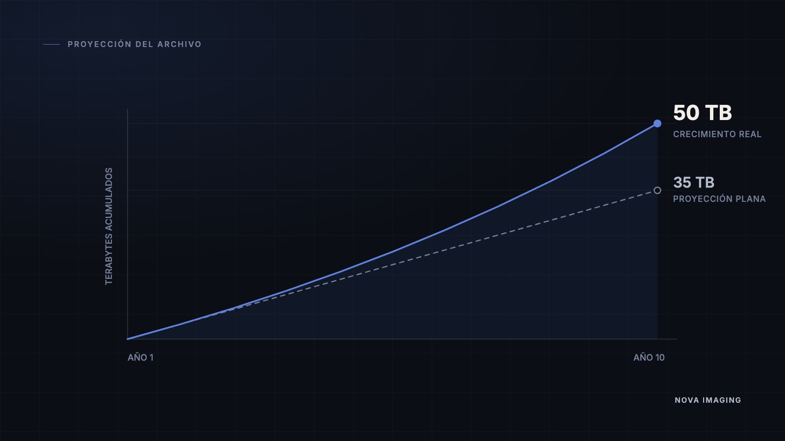
  2. Storage that grows without surprises.Lossless compression and automatic tiering · predictable costs
  3. Automatic worklist prioritization.By criticality, modality and payer · lists configurable per shift
  4. Reports the AI reviews.Contrasts what was written against what was marked · alerts, never blocks
50M+

Medical images processed each year, quickly and accurately.

30%

Lower storage costs through image compression.

1M+

Patients reach their results through our secure portal.

70%

Fewer administrative errors with RIS, PACS and HIS integration.

Platform

The whole life of a study, in one system

WorklistReading
Prioritize critical
ModalitiesPrioritiesPayersCreate list
All7My studies0Mammography0Columns
ModPatient · studyPrioTime
DX
Urgent
Gómez M., J.C.
Chest PA · EX-0001
14m
MR
Rodríguez P., M.
Brain MRI · MR-0156
HIGH2d
US
López S., C.
Renal ultrasound · US-0234
HIGH2d
CT
Díaz G., F.
Abdominal CT · CT-0456
MED5d
MG
Torres N., P.
Bilateral mammogram · MG-0678
LOW7d
7 pending · view "All"Shared · night shift

Intelligent worklist

The work queue sorted by what is actually urgent, not by order of arrival.

  • Prioritizes by clinical urgency, modality, payer contract and SLA
  • Fully customizable lists, shareable across shifts
  • Reads the order data (HL7/FHIR) — never the image
portal.novaimaging.co/viewer
Synced
RID · XXXXXXX
CT CHEST-ABDOMEN
01-Jul-2026 · 02:19:12
AW:360 L:60181/362
AID · XXXXXXX
CT CHEST-ABDOMEN
01-Jul-2026 · 02:19:12
HW:360 L:60253/512
RID · XXXXXXX
CT CHEST-ABDOMEN
01-Jul-2026 · 02:19:12
HW:360 L:60257/512
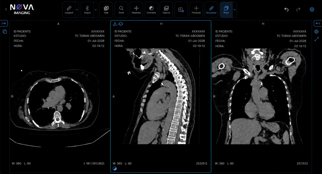
NOVA Imaging viewer: chest-abdomen CT in axial, sagittal and coronal planes

DICOM viewer in the browser

Diagnostic reading without installing a client on every workstation.

  • Windowing, measurements and comparison with prior studies
  • MPR, MIP and GPU-rendered volume
  • Built on Cornerstone3D · OHIF, zero-footprint
Chest CT · templateSigned

Findings

Solid nodule in the right upper lobe measuring 14 mm, with regular borders. No mediastinal lymphadenopathy.

Impression

Solitary pulmonary nodule. CT follow-up is suggested.

R91.8 · Abnormal lung findingSearch ICD-10

Structured reporting

From dictation to signature without leaving the same system.

  • Templates per study type, with clinical dictation and macros
  • Digital signature with version control
  • ICD-10 diagnosis lookup tied to the impression
HIS / EMRMedical recordPatient portalStudies and reportsPhysician portalReferrersERPBillingNOVA Enterprise ImagingFHIR · HL7 · DICOM

Interoperability

Connects with what your institution already has, without replacing it.

  • HL7 v2 (ADT · ORM · ORU) and FHIR R4 over a REST API
  • DICOM, DICOMweb (WADO · QIDO · STOW) and Modality Worklist
  • IHE profiles: XDS-I, PIX/PDQ and SWF
portal.novaimaging.co
EMElena MarínPatient portal · 3 studies
MRRight knee MRIJul 12, 2026View study
CTAbdominal CTMar 3, 2026View study
USThyroid ultrasoundNov 18, 2025View study

Patient and physician portals

Results and images through a secure link. No CDs, no film.

  • The patient opens the report and images whenever they need them
  • The referring physician reaches the studies of the patients they referred
  • An AI agent explains the report to the patient in plain language
quality dashboard
Risk level
Low10%Medium72.5%High17.5%
By modality
CTUSMREPDX
Summary by physician
M. Torres Vega98.9Excellent
R. Salas Nieto100Excellent

AI quality control

A second pair of eyes over reports that are already signed.

  • Audits closed reports in batches and flags what deviates
  • Compares the report against the measurements and findings in the study
  • Feeds the service quality program, not a ranking
report editor · AI
Findings

Focal lesion in the left kidney,

of cystic appearance, measuring 42 mm

with well-defined borders.

AI detectionLive
Laterality

“Left” does not match the R marker on the image.

Classification

Renal cystic lesion with no Bosniak category assigned.

Inconsistency detection

While the radiologist writes, not after delivery.

  • Flags a laterality or a measurement that does not match the image
  • Runs inline, without interrupting dictation
  • It alerts and suggests: the clinical decision is always the radiologist’s
Archive · 10-year projectionAutomatic
Fast0 – 90 days

Studies being read and recent comparisons

Standard90 days – 2 years

Occasional access from the portal

Cold archive2 – 10+ years

Legal retention of the medical record

Storage and deployment

Predictable cost over ten years and the deployment model that suits you.

  • Compression, deduplication and automatic archive tiering
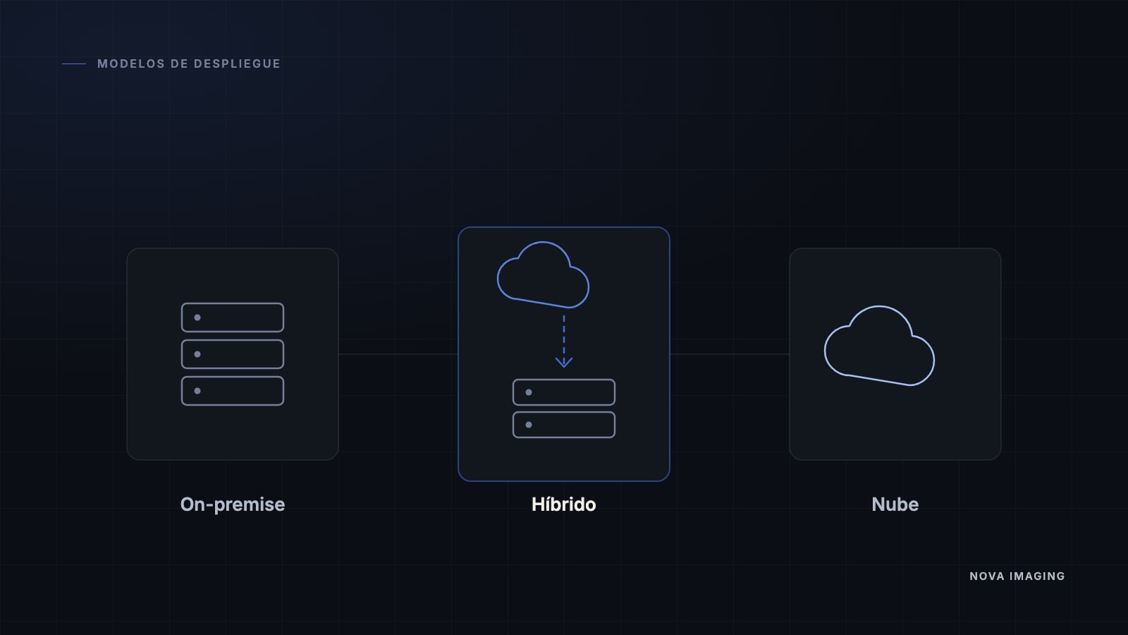
  • Fully cloud, on-premise or hybrid
  • Backup, disaster recovery and migration of the legacy DICOM archive
See the full platform
The NOVA Imaging ecosystem

One system. Every part of the radiology workflow.

Four modules designed to work together — from dictation to the patient portals — all on the same platform.

01 / 04

Dictation

State-of-the-art clinical dictation

Medical transcription with radiology vocabulary in Spanish, English and Portuguese. Voice commands for macros and signing, and fluid dictation of measurements and findings.

Dragon Medical OneSpeechKit

Listening · en-US

Template · Chest CT without contrast

⌘.
00:24A subpleural nodule is identified measuring00:27forty two point seven millimeters→ 42.7 mm
02 / 04

Worklist

Fully customizable work lists

Each radiologist defines their own columns, priorities and saved filters. Shareable across teams and shifts.

WorklistReading
Prioritize critical
ModalitiesPrioritiesPayersCreate list
All7My studies0Mammography0Columns
ModPatient · studyPrioTime
DX
Urgent
Gómez M., J.C.
Chest PA · EX-0001
14m
MR
Rodríguez P., M.
Brain MRI · MR-0156
HIGH2d
US
López S., C.
Renal ultrasound · US-0234
HIGH2d
CT
Díaz G., F.
Abdominal CT · CT-0456
MED5d
MG
Torres N., P.
Bilateral mammogram · MG-0678
LOW7d
7 pending · view "All"Shared · night shift
03 / 04

Portals

Patient and referrer portals

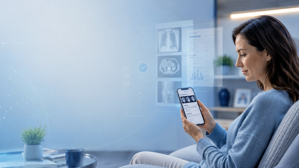
View images, share studies, and an AI agent that explains reports to patients in plain language.

AI agent
EM
Elena M.
Patient portal
2 studies
CT · Apr 183 series

NOVA explains · in plain words

"Your study shows an area the doctor wants to look at closely at an upcoming visit. It is not urgent."
Link · 72hPatientReferrer
04 / 04

Architecture

Fully web & flexible

Cloud-native, with on-premise or hybrid deployments to suit your client. Scales without friction and without reinstalling heavy clients.

Cloud-native
AWS · GCP · Azure
On-premise
In your data center
Hybrid
Best of both
Operational intelligence

The AI that respects the radiologist

At NOVA Imaging, the AI never blocks the read and never diagnoses for the radiologist. It acts at three moments: before the read it prioritizes the queue, during dictation it flags without slowing anyone down, and after the signature it audits in batches. The clinical decision is always the human’s.

Before the read · in real time

Prioritization of critical cases

NOVA Imaging reads the clinical data —not the images— and reorders the worklist before the radiologist opens the next study. What is urgent rises to the top on its own.

The AI can:

  • Read the clinical data (HL7 · FHIR), never the image in order to diagnose
  • Reorder the worklist in milliseconds, in the background
  • Prioritize critical cases by severity and by your rules
How it works
intelligent worklist · AI prioritization

Clinical data (not the image)

HL7 ADT · admitted with chest pain
FHIR · elevated troponin (3.1 ng/mL)
Order · Chest CT · "rule out PE"
History · D-dimer > 2000
ER triage · ESI-2 priority
Score 96/100 → probable PE. Raise to STAT.

Worklist · reorderedlive

STAT
García R., M.
CT · Chest
96
TODAY
Vega A., R.
US · Carotid
41
TODAY
Ramírez K., L.
XR · Chest
34
48h
Núñez T., S.
MR · Lumbar
18
24h
Soto V., M.
MR · Knee
12
Median latency: 180 msprocessing queue
After the signature · in batches

Report quality control

This layer is different from the inline alerts: it runs after the signature, in batches, over the already closed report. It flags serious anatomical and laterality inconsistencies for review the next day, without touching the live workflow.

The AI can:

  • Review signed reports in batches, after the live workflow
  • Contrast laterality and anatomy in the text against what was marked in the study
  • Flag only what is serious, without slowing the radiologist’s reading
Model and validation
quality control · batch audit

Signed reports · day247 today

Batch control
08091011121314151617181920212223

Batch findings · review next day3 of 247 · 1.2%

R-2289Laterality · "left" vs R markeralta
R-2304Anatomical mismatch · organ referencedalta
R-2317Incomplete template · findingsmedia
False positives: < 0.3%overnight batch · 02:00
Clinical differentiators

What no generic RIS/PACS ships with

quality dashboard
Risk level
Low10%Medium72.5%High17.5%
By modality
CTUSMREPDX
Summary by physician
M. Torres Vega98.9Excellent
R. Salas Nieto100Excellent

AI report quality control

A second pair of eyes before signing

NOVA Imaging compares the report text against the measurements and findings marked in the viewer, and flags omissions or discrepancies before the report leaves the system.

report editor · AI
Findings

Focal lesion in the left kidney,

of cystic appearance, measuring 42 mm

with well-defined borders.

AI detectionLive
Laterality

“Left” does not match the R marker on the image.

Classification

Renal cystic lesion with no Bosniak category assigned.

Real-time inconsistency detection

Alerts while the radiologist writes

If a measurement or laterality in the text does not match what is marked on the images, NOVA Imaging highlights it instantly — not after the signature.

portal.novaimaging.co
EMElena MarínPatient portal · 3 studies
MRRight knee MRIJul 12, 2026View study
CTAbdominal CTMar 3, 2026View study
USThyroid ultrasoundNov 18, 2025View study

Patient portal

Results within reach, no phone calls, no CDs

The patient and the referring physician open the report and the images from a secure portal, with access links controlled per study.

portal.novaimaging.co/viewer
Synced
RID · XXXXXXX
CT CHEST-ABDOMEN
01-Jul-2026 · 02:19:12
AW:360 L:60181/362
AID · XXXXXXX
CT CHEST-ABDOMEN
01-Jul-2026 · 02:19:12
HW:360 L:60253/512
RID · XXXXXXX
CT CHEST-ABDOMEN
01-Jul-2026 · 02:19:12
HW:360 L:60257/512
NOVA Imaging viewer: chest-abdomen CT in axial, sagittal and coronal planes

Zero-footprint visualization

An advanced workstation, not just a viewer

MPR, MIP, volume rendering and hanging protocols configurable per study — diagnostic workstation tools that run in the browser, with no client installed on the machine.

Who already uses it

Colombian institutions running on the platform today.

What the medical directors who implemented it say

The integration between NOVA Imaging and our Electronic Health Record improved our processes and the quality of care. We want to highlight the team’s professionalism and technical capability in delivering a robust, seamless RIS/PACS solution.
Dr. David Ricardo VélezDirector of Technology and Data Science, Clínica Angiosur
NOVA Enterprise Imaging streamlined how we manage studies, with a platform that is fast, secure and fully integrated into our processes. Their technical support has been key to improving care and institutional efficiency.
Dra. Mónica María LópezGeneral Manager, IPS Ramiro Correa Restrepo S.A.S
NOVA Imaging offers a versatile, intuitive and secure RIS/PACS, with ERP/HIS integration and constant support. Its impact on healthcare institutions is tangible: a robust and transformative solution.
Juan Camilo SaldarriagaCommercial Director, Createc
Impact calculator

How much physician time does your institution get back?

Adjust your monthly volume and your average time per report. We estimate the physician hours recovered per year.

Conservative estimate
Monthly study volume8,000 studies / month
50010k20k30k40k
Average time per report11 min
310152030

How we calculate it. Clinical dictation and macros recover ≈22% of reporting time; prioritization and a unified queue save ≈1.5 min per study in waiting and context switching. Validated with 6 pilot institutions.

Physician hours recovered / year

6,276hrs

Equivalent to3.5 full-time physicians
Time saved per study3.9 min
Monthly savings523 hrs
Technology backing

Built on first-rate infrastructure

NOVA Imaging takes part in the partner programs of Google, Anthropic and Cloudflare, and runs on their compute, network and AI model infrastructure.

GoogleGoogle for Startups
AnthropicClaude Partner Network
CloudflareCloudflare for Startups

That backing turns into a faster, safer and smarter product for your center.

Shall we talk about how this would look at your center?

We will show you the platform running with your institution’s real workflow and answer your questions about migrating the existing archive.